

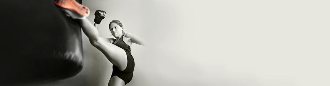


Hip Impingement (Femoroacetabular Impingement Or FAI)
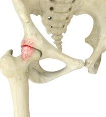
FAI is a condition affecting the hip joint articulation. It is characterized by an abnormal contact between the femoral head (hip ball) and the acetabular rim (hip socket) leading to eventual damage of the labrum and articular cartilage that lines the bone surface. This may in turn lead to arthritis of the hip joint.
The labrum (which translates into “lip”) is a cartilage ring that lines the hip socket similar to the labrum of the shoulder joint. It serves several functions including: 1) to ensure a smooth and even contact pressure between the hip head and acetabulum, 2) keeping the head from dislocating out of the acetabulum, and 3) dispersing the synovial fluid that bathes the articular cartilage with nutrients. The smooth articular cartilage that lines the bones in our joints serves to allow joints to move without causing excess friction and damage.
In the setting of FAI, a prominence of abnormal bone on the femoral head or the acetabulum causes the two bones to collide through an arc of motion. There are generally three types of impingement: CAM impingement (prominence on femoral neck), PINCER impingement (prominence of acetabular socket) and mixed impingement. These bone prominence can form from a variety of causes. Patients with painful FAI develop groin pain with hip motion. As the head rotates in the acetabulum, the labrum may become pinched and torn between the head and acetabulum.
FAI can affect all age groups ranging from the early teens to throughout adult life. There is significant research recognizing that this disorder can lead to eventual hip osteoarthritis, even in the early adult years. Certain activities that involve repetitive hip motion may increase the frequency of this bone contact (ie. kicking sports, ballet, hockey, golf).
FAI is diagnosed by a careful physical examination by your surgeon in conjunction with diagnostic studies such as x-rays, MRI scans, and CT scans. Some patients may be advised to undergo a diagnostic and possible therapeutic injection of the hip joint. This can be performed using fluoroscopy (x-ray) or ultrasound to guide the injection into the joint space. This is often best performed by your treating surgeon and can be performed in the office with minimal discomfort.
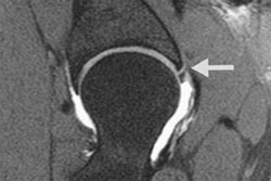
MRI scan Showing Labrum Tear
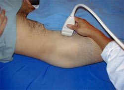
Ultrasound Scan of Hip
There are several treatment strategies for FAI. Most patients should undergo physical therapy of the hip to see if this alleviates pain and restores joint motion. Some patients may be advised to undergo a diagnostic and possible therapeutic injection of the hip. This can be performed using fluoroscopy (x-ray) or ultrasound to guide the injection into the joint space. Hip arthroscopy may help to address the labrum tear in the hip joint. The tear can be repaired or debrided using this minimally invasive approach. If the labrum can be repaired, sutures are placed to re-appose the labrum to the acetabulum to allow healing to the bone. The labrum may need to be temporarily released from the acetabulum in some cases to allow for complete impingement resection. The bony prominences causing the impingement of the femoral neck and acetabulum can be resected. This can improve the hip pain and restore the hip range of motion. The most common location of these prominences makes them accessible with standard hip arthroscopic techniques. In certain conditions, a formal open approach must be performed. At the conclusion of the case, the hip capsule tissues may be sutured closed. In patient with subtle instability, the capsule may be plicated to reduce the volume of the hip joint.
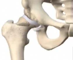
Hip Pincer Lesion with Labrum Tear
Twenty-two year-old soccer player with progressive hip pain over the past 2 years. The patient originally injured his hip playing during a free-kick and has tried conservative management including physical therapy, anti-inflammatories, and rest. Radiographs demonstrated a large well-corticated bony prominence adjacent to the anterolateral acetabulum labrum extending towards the anterior inferior iliac spine. MRI scan showed a tear of the anterolateral labrum. The patient underwent an arthroscopic resection of the bony impingement lesion as well as fixation of the labrum. He remains pain-free and has had no hip residual pain.
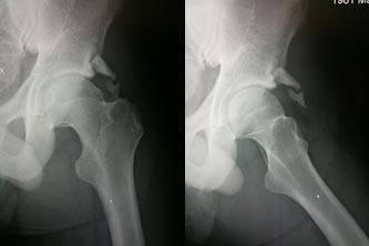
Prior to Resection of the Impingement Lesion
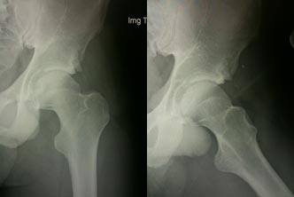
After Arthroscopic Impingement Resection