







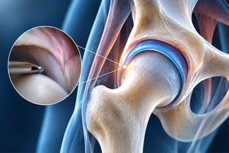
Hip arthroscopy looks simple from the outside: a camera, small incisions, and a joint repaired through portals the width of a pencil. Inside the joint, it is one of the most technically demanding operations in orthopedics. The hip is deep, tight, and unforgiving. There is little room for error, the anatomy varies from patient to patient, and the difference between a good result and a disappointing one often comes down to diagnosis — not just technique.
Dr. J. Pieter Hommen has performed hip arthroscopy for more than 20 years, through the period when the operation grew from a niche diagnostic tool into a standard treatment for a wide range of hip problems. That length of experience matters, not because time alone guarantees a result, but because it changes how a surgeon sees the whole problem: who should have surgery, who should not, and what needs to be fixed — and what needs to be left alone — once inside.
How hip arthroscopy has advanced
Early hip arthroscopy was limited by access, instruments, and imaging. Surgeons could see less, repair less, and protect the joint less reliably. Over the past two decades, several advances have changed that:
- Better traction and positioning, with careful attention to limiting traction time and protecting nerves and skin
- Improved cameras, fluid management, and instruments designed specifically for the hip
- A clearer understanding of the capsule, the strong ligament envelope around the hip, and why it usually needs to be repaired or closed after surgery
- Stronger, smaller anchors and suture techniques for labral repair and reconstruction
- Peritrochanteric endoscopy — arthroscopy outside the joint — for gluteal tendon tears and other problems around the outside of the hip
- Better preoperative imaging, including MRI arthrography and 3D assessment of hip shape, so the surgical plan matches the anatomy before the first incision
The operation is still minimally invasive in terms of incision size. It is not minor surgery. The joint must be distracted — gently pulled apart — to work inside it, and the surgeon works in compartments: the central compartment inside the socket, the peripheral compartment around the neck of the femur, and the peritrochanteric space outside the joint.
Why it takes years to master
Research on the hip arthroscopy learning curve is unusually blunt. Studies have found that operative and traction times fall significantly after roughly 70 to 110 cases, and one large database study of more than 8,000 hip arthroscopies found that the risk of needing further surgery did not fall appreciably until a surgeon had performed at least 389 cases. That finding is not about speed. It reflects judgment: recognizing the right diagnosis, selecting the right patient, and completing the full correction the first time.
Mastery, in this operation, is really three skills at once. First, diagnosis — matching groin pain, catching, or instability to a specific structure on examination and imaging, and ruling out the spine, sacroiliac joint, sports hernia, and other sources of hip-area pain. Second, technical execution in a tight space, where an instrument placed a few millimeters off can injure cartilage. Third, restraint — knowing when a labral tear on MRI is not the cause of pain, and when arthritis or dysplasia (a shallow socket) means arthroscopy alone is the wrong operation.
Strong indications are the foundation of happy results
The happiest results in hip arthroscopy share a pattern, and it starts before surgery. The patient has a clear mechanical problem, the examination reproduces the pain, the imaging matches the examination, conservative treatment has been tried, and the joint itself is not already worn out by advanced arthritis.
Indications Dr. Hommen has treated over more than two decades include:
- Femoroacetabular impingement (FAI), where the shape of the ball or socket causes abnormal contact during movement — cam, pincer, and mixed patterns
- Labral repair, restoring the ring of cartilage that seals the joint and contributes to stability
- Labral reconstruction, when the native labrum is too damaged or deficient to repair, using graft tissue to rebuild the seal
- Gluteus medius and minimus repairs, for tears on the outside of the hip that cause lateral pain, limping, and weakness
- Hamstring repairs, particularly proximal hamstring tears at the sitting bone
- Iliopsoas tendon release, including painful iliopsoas impingement after hip replacement surgery, when the tendon rubs against the replacement cup
- Chondral lesions, or areas of damaged cartilage inside the joint
- Hip dislocations and instability, loose bodies, and other mechanical problems inside the joint
No list guarantees a result. A labral tear, for example, can be present without causing pain, and fixing it will not help pain that comes from somewhere else. That is why the indication — the reason for surgery, proven as carefully as possible — matters more than the size of the incision.
What patients should take from 20 years of experience
Experience does not make hip arthroscopy right for everyone. It makes the conversation more honest. A surgeon who has seen both excellent results and disappointing ones is more likely to say, plainly: this problem fits arthroscopy well; this problem needs a different operation, such as a periacetabular osteotomy for dysplasia or a hip replacement for advanced arthritis; or this problem needs more time, therapy, and diagnosis before any operation.
If you are considering hip arthroscopy, ask about the specific diagnosis, what will be repaired, what the capsule plan is, what rehabilitation looks like, and what the realistic risks and alternatives are. The best operations start with the strongest indications.
This post is for education only. It is not medical advice and does not promise a specific result. Every hip is different, and the right treatment depends on an in-person evaluation, examination, and imaging.
Selected references: Learning-curve studies in hip arthroscopy, including prospective evaluation with 2-year outcomes in FAI and a scoping review of arthroscopic learning curves; large database analysis of surgeon volume and reoperation after hip arthroscopy.